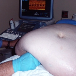
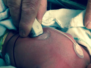
My first boss in this business taught me to see it, learn it, do it and then teach it. I believe that Dr. William Rashkind would have approved of this website. It is dedicated to him, Drs. Sidney Friedman, William Norwood, Paul Weinberg, Henry Wagner and Victoria Lee Vetter at CHOP and then to Drs. Bolognese, Librizzi and Weiner and to Vincenzo Berghella at Pennsy and Jefferson as well as to those fetuses and newborn babies I have met through the application of diagnostic ultrasound. The website is established at the request and insistence of various students, sonographers, nurses and colleagues from around the world with whom I have had the pleasure to work. Most of these anonymized studies are from my personal collections while a number were donated by national and international colleagues. I will include more of your studies as they become available.
The premise of this website is that as structural congenital heart disease is well known to be the most common, the most associated with genetic syndromes and the most lethal of all congenital anomalies and malformations, CHD should be recognized on routine obstetrical ultrasound examinations as variations in position, size, function, rhythm, and chamber and vessel size proportion. In addition, the neonatologist should be able to utilize ultrasound to evaluate the neonatal heart for structure and function.
We will attempt to demonstrate normal cardiovascular anatomy and physiology and then the wide variety of structural and functional heart variations that can be detected with ultrasound. Simple rules of imaging, following the segmental approach to analysis of the veins, valves,chambers, and arteries are shown and described in still and in real time ultrasound imaging. A future search button on the home page will allow the site visitor to find videos of specific lesions as well as papers published on that topic.
This website offers an overview of imaging seen by ultrasound of the fetal or neonatal heart and is not intended to replace formal education in radiographic, obstetric or pediatric cardiac ultrasound. These images do not represent or constitute professional diagnostic advice. A bibliography of current and recent guidelines by national and international medical societies is incorporated in the indices of published papers relating to fetal and neonatal cardiovascular ultrasound and pediatric cardiology.
All interested readers are invited to send fetal and newborn heart imaging studies for inclusion in this website to PERINATALECHOIMAGING.COM. All submissions will be converted to our de-identified format and annotated. There is no external financial support for this website. Users of this website are invited to support this effort and donate to its upkeep. valves, chambers, and arteries are shown and described in still and in real time ultrasound imaging. A search button on the home page will allow the site visitor to find videos of specific lesions as well as papers published on that topic.
This website offers an overview of imaging seen by ultrasound of the fetal or neonatal heart and is not intended to replace formal education in radiographic, obstetric or pediatric cardiac ultrasound. These images do not represent or constitute professional diagnostic advice. A bibliography of current and recent guidelines by national and international medical societies is incorporated in the indices of published papers relating to fetal and neonatal cardiovascular ultrasound and pediatric cardiology.
All interested readers are invited to send fetal and newborn heart imaging studies for inclusion in this website to PERINATALECHOIMAGING.COM. All submissions will be converted to our de-identified format and annotated. There is no external financial support for this website. Users of this website are invited to support this effort and donate to its upkeep.
Quick evaluation for cause
Always look at the right and left outflow tracts
for position, flow direction, number of vessels
and variations in the 3 Vessel Views
for position, flow direction, number of vessels
and variations in the 3 Vessel Views

